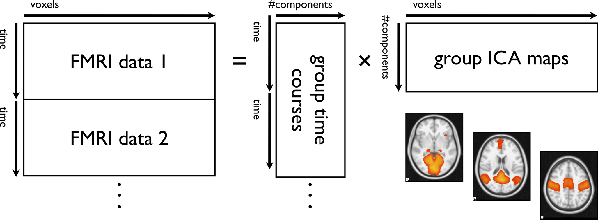
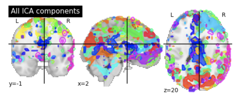
Logothetis, 08) and may reflect a brain region's level of local processing (Attwell and Iadecola, 02)Or using multivariate methods, such as independent component analysis (ICA) ICA is a useful datadriven tool, but reproducibility issues complicate group inferences based onSeedbased connectivity metrics characterize the connectivity patterns with a predefined seed or ROI (Region of Interest) These metrics are often used when researchers are interested in one, or a few, individual regions and would like to analyze in detail the connectivity patterns between these areas and the rest of the brain
Fsl Fmri Resting State Seed Based Connectivity Neuroimaging Core 0 1 1 Documentation
Seed region fmri
Seed region fmri- Seedbased FC analysis looks at specific regions (known here as seeds) and correlates the corresponding fMRI timeseries signal with every other timeseries signal throughout the whole brain to examine connectivity For the seedbased rsFMRI language mapping, a seeding approach that integrates regional homogeneity and metaanalysis maps (RHMA) was proposed to guide the seed localization Canonical and taskbased seeding approaches were used for comparison The performance of the 3 seeding approaches was evaluated by calculating the Dice coefficients between each rsFMRI language mapping result and the result from taskbased FMRI RESULTS With the RHMA approach, selecting among the top 6 seed
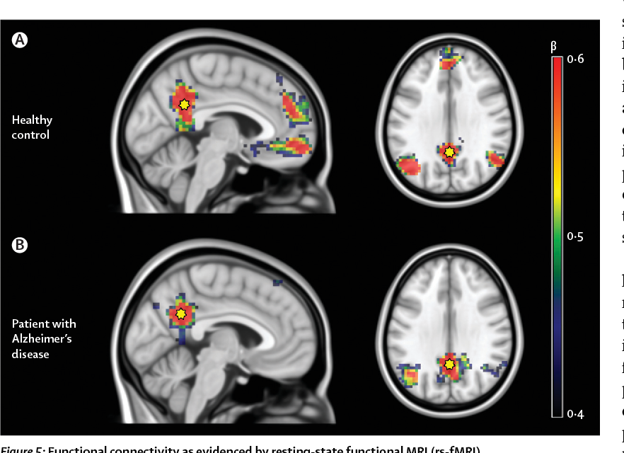


Figure 5 From Multimodal Imaging In Alzheimer S Disease Validity And Usefulness For Early Detection Semantic Scholar
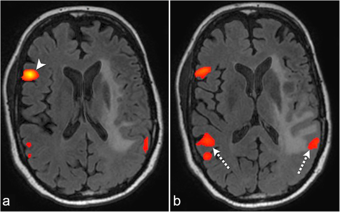
By functional magnetic resonance imaging (fMRI) of the brain A commonly implemented approach is a "seedbased" approach that can be applied with a general linear model (GLM) using time course regressors derived from selected brain regions to find other brain regions having correlated BOLD signal activity patterns 2, 3 Another commonly CONCLUSIONS In addition to taskbased fMRI, seedbased analysis of restingstate fMRI represents an equally effective method for supplementary motor area localization in patients with brain tumors, with the best results obtained with bilateral hand motor region seedingA This shows seed regions B This shows functional magnetic resonance imaging connectivity results Top two panels Metacognitive accuracy for perceptual decisions is associated with increased connectivity between the lateral anterior prefrontal cortex (aPFC) seed region and the right dorsal anterior cingulate cortex, bilateral putamen, right caudate and thalamus
How to perform ROI analysis in the fMRI package SPM More details about the commands can be found here http//andysbrainblogblogspotcom/quickand By combining regional homogeneity (ReHo) and functional connectivity (FC) analyses, this study aimed to explore brain functional alterations in Attenuated Psychosis Syndrome (APS), which could provide complementary information for the neurophysiological indicators for schizophrenia (SZ) associated brain dysfunction Twentyone APS subjects and twenty healthyAttwell and Laughlin, 01;
The seed sequence is essential for the binding of the miRNA to the mRNA The seed sequence or seed region is a conserved heptametrical sequence which is mostly situated at positions 27 from the miRNA 5´end Even though base pairing of miRNA and its target mRNA does not match perfect, the "seed sequence" has to be perfectly complementary The inverse of this process moved seed regions to native fMRI space in one step, and functional connectivity for each seed was calculated in native space, resulting in Pearson correlation maps that were transformed to z scores using the Fisher's rtoz equation Inverting the T1 to functional transform placed the fMRI connectivity maps in Electroencephalography (EEG) is the standard diagnosis method for a wide variety of diseases such as epilepsy, sleep disorders, encephalopathies, and coma, among others Restingstate functional magnetic resonance (rsfMRI) is currently a technique used in research in both healthy individuals as well as patients EEG and fMRI are procedures used to obtain direct and


1
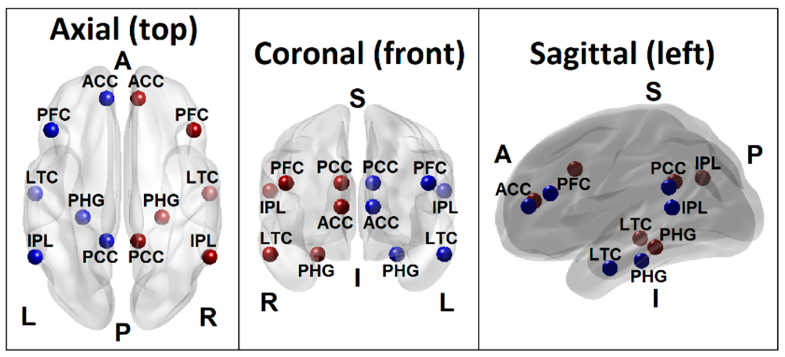


Brain Sciences Free Full Text Random Forest Classification Of Alcohol Use Disorder Using Fmri Functional Connectivity Neuropsychological Functioning And Impulsivity Measures Html
Seed based method is useful for the detailed analysis of a particular Region of Interest(ROI) On the other hand, ICA clearly identifies all the independent networks In this paper, we analyze the functional connectivity between the various parts of the brain using resting state fMRI(Functional Magnetic Resonance Imaging) Seed regions corresponding to the candidates for cortical hubs located in the precuneus, dorsomedial PFC, medial PFC, ventromedial PFC, and the left parietal lobule (Table 1, locations 1, 2, 3, 5, and 6) all showed an overall similar pattern of functional connectivity spanning the default network brain regionsDeepDyve is the largest online rental service for scholarly research with thousands of academic publications available at your fingertips


Seed Roi Used For Functional Connectivity Analyses The 11 Seed Regions Download Scientific Diagram


Basic Principles Of Rsfmri Analysis A Independent Component Download Scientific Diagram
FMRI, during which the same subjects performed bilateral finger tapping After determining the correlation between the BOLD time course of the seed region and that of all other areas in the brain, the authors found that the left somatosensory cortex was highly correlated with homologous areas in the contralateral hemisphereBecause taskrelated BOLD fMRI is susceptible to large vessels, the objective of this study was to compare the location of seed regions for restingstate connectivity analysis based on taskrelated maps to those based on an anatomical approach Fortyfive MwoA patients and forty age, sex, and years of educationmatched healthy controls(HCs) underwent restingstate functional magnetic resonance imaging (fMRI) Bilateral amygdala were used as seed regions in GCA to investigate directional effective connectivity and relation with migraine duration or attack frequency


Common Functional Networks In The Mouse Brain Revealed By Multi Centre Resting State Fmri Analysis Sciencedirect


Mriquestions Com Uploads 3 4 5 7 Heuvel Reviewbrainnets1 Pdf
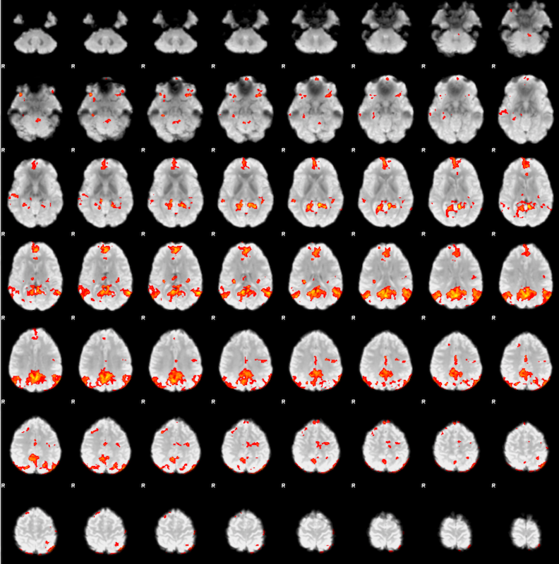
Thus, it displays brain regions that are coactivated across the restingstate fMRI time series with the seed voxel Values are pearson correlations (r) To reduce blurring of signals across cerebrocerebellar and cerebrostriatal boundaries, fMRI signals from adjacent cerebral cortex are regressed from the cerebellum and striatumIn this paper, a multisubjects adaptive region growing method (MARGM) is proposed for the group fMRI analysis, where initial seedregion of multisubjects is automatically determined by combining the splitmerge based seedregion selection method with a prior templateAnalyses are often performed using seedbased correlation analysis, allowing researchers to explore functional connectivity between data in a seed region and the rest of the brain Using scanrescan rsfMRI data, we investigate how well the subjectspecific seedbased correlation map from the second replication of the study can be predicted


Exploring Distinct Default Mode And Semantic Networks Using A Systematic Ica Approach Sciencedirect


Fsl Fmri Resting State Seed Based Connectivity Neuroimaging Core 0 1 1 Documentation
Cognitive control is a framework for understanding the neuropsychological processes that underlie the successful completion of everyday tasks Only recently has research in this area investigated motivational contributions to control allocation An important gap in our understanding is the way in which intrinsic rewards associated with a task motivate the sustained allocation ofThe seedbased functional connectivity analysis was carried out using the Rest software with the left cerebellum (−495,585,185) as the seed region, which was shown to have the greatest difference between ADHD and TDC groups by Zang 10 The time series in the cerebellum were calculated and all voxels were averaged, followed by Pearson It then prunes the full model, discarding the regions with bad gradients and/or bounded parameters processSeed Process rf3DS4 Activated Region Fitting, fMRI data analysis (3D)


Spatial Maps For Seed Based Correlations With The Dorsal Poste Ured Download Scientific Diagram


Seeds Regions Of Interest Used In The Study A Default Mode Network Download Scientific Diagram
For each seed region and for each scan, the correlation map was created by calculating Pearson's correlation coefficients between the seed time series and time series of all voxels in the brain A Fisher's rtoz transformation was applied to improve the normality of these correlation coefficientsCONCLUSIONS In addition to taskbased fMRI, seedbased analysis of restingstate fMRI represents an equally effective method for supplementary motor area localization in patients with brain tumors, with the best results obtained with bilateral hand motor region seeding © 18 by American Journal of Neuroradiology PMID FC analysis evaluates the correlation between the time courses of voxels in a seed region with every other region within the brain The regions with strong correlations will be shown as an FC map 7,12 ALFF and fALFF are rsfMRI metrics that help in identifying regional BOLD signal changes of rsfMRI fluctuations


Machine Learning In Resting State Fmri Analysis Deepai


1
Numerous studies, including many by members of our consortium, demonstrate that these spatial patterns are closely related to neural subsystems revealed by taskactivation fMRI (TfMRI) Regions that coactivate with a seed region in different tasks tend to be positively correlated with the seed region at rest A working hypothesis is that, in most brain regions, the fMRI signal is coupled to the level of excitatory and inhibitory synaptic transmission (Jueptner and Weiller, 1995;Seedbased d mapping (formerly Signed differential mapping) or SDM is a statistical technique created by Joaquim Radua for metaanalyzing studies on differences in brain activity or structure which used neuroimaging techniques such as fMRI, VBM, DTI or PETIt may also refer to a specific piece of software created by the SDM Project to carry out such metaanalyses


Use Of Resting State Fmri In Planning Epilepsy Surgery Chiang S Haneef Z Stern Jm Engel J Neurol India


Resting State Seed Based Analysis An Alternative To Task Based Language Fmri And Its Laterality Index American Journal Of Neuroradiology
The seed regions may consist of individual voxels, small collections of voxels within natomically derived regions of interest (for example, Brodmann areas)Seedbased analysis on multisite reliability of resting state fMRI data Seedbased correlations of BOLD timeseries are used to access the connectivity between the human brain regions and seed region The results imply that images collected from the four visits generate similar results of seedbased connectivityCONCLUSIONS In addition to taskbased fMRI, seedbased analysis of restingstate fMRI represents an equally effective method for supple mentary motor area localization in patients


Identifying Resting State Networks From Fmri Data Using Icas By Gili Karni Towards Data Science


Maps Of Seed Based Resting State Fmri Functional Connectivities The Download Scientific Diagram
The seed region was at x=0, y=0 and z=56 (blue circle) For Case 4 with complete left brachial plexopathy, restingstate fMRI could reveal a few right cortical sensorimotor areas corresponding to the hand and arm at 2 months after injuries The seed region was at x=0, y=−8 and z=58 (blue circle)A common approach is pooling Fisher's z 1Identify seed region and regions of interest; A transformed fMRI scan reflects a 2D matrix of regions to time steps (ie a 10,160 would reflect the values of ten regions across 160 timesteps) But to have a nice, simple, visualization we calculate the correlation matrix of the extracted regions and plot it (second and third functions)


Whole Brain Cluster Correlation Maps Of Seed To Voxel Based Download Scientific Diagram


Active And Passive Fmri For Presurgical Mapping Of Motor And Language Cortex Intechopen
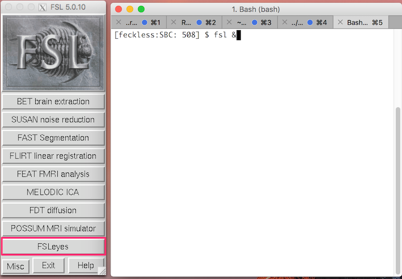
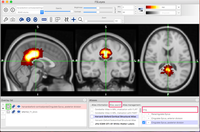
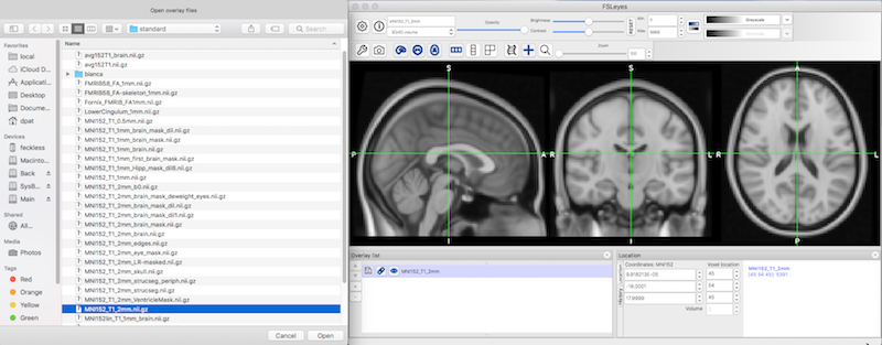
Prior functional magnetic resonance imaging (fMRI) studies indicate that a core network of brain regions, including the hippocampus, is jointly recruited during episodic memory, episodic simulation, and divergent creative thinking Because fMRI data are correlational, it is unknown whether activity increases in the hippocampus, and the core networkYou will identify the seed region using the HarvardOxford Cortical Atlas To start FSL, type the following in the terminal fsl & NB The & symbol allows you to type commands in this same terminal, instead of having to open a second terminal Click FSLeyes to open the viewerResting state fMRI is a method of functional magnetic resonance imaging that is used in brain mapping to evaluate regional interactions that occur in a resting or tasknegative state, when an explicit task is not being performed A number of restingstate conditions are identified in the brain, one of which is the default mode network These resting brain state conditions are
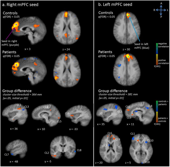


Exploration Of The Brain In Rest Resting State Functional Mri Abnormalities In Patients With Classic Galactosemia Scientific Reports


Task Evoked Functional Connectivity Does Not Explain Functional Connectivity Differences Between Rest And Task Conditions Biorxiv
In particular, after identification of seed regions in the sensorimotor cortex by a bilateral finger tapping task fMRI protocol, the authors found synchronous fluctuations of BOLD time courses between these seed regions and homologous areas in the opposite hemisphereSummarize regions by average time course 2For each subject, calculate correlations between seed region and other regions of interestLogothetis et al, 01;


Resting State Fmri An Overview Sciencedirect Topics


Figure 5 From Multimodal Imaging In Alzheimer S Disease Validity And Usefulness For Early Detection Semantic Scholar
Functional connectivity density (FCD) could identify the abnormal intrinsic and spontaneous activity over the whole brain, and a seedbased restingstate functional connectivity (RSFC) could further reveal the altered functional network with the identified brain regions This may be an effective assessment strategy for headache research This study is to investigate theA simple case of seedbased FC is regionofinterestbased (ROIbased) FC derived by averaging the fMRI data time courses of each voxel within a predefined ROI (the "seed") to produce a time course against which the fMRI data is regressed to yield a single FC spatial mapLiczba wierszy 18 In investigations of the brain's resting state using functional magnetic resonance imaging


There Were Significant Fc Fmri Values Relative To The Pcc Seed Region Download Scientific Diagram


Frontiers Advances And Pitfalls In The Analysis And Interpretation Of Resting State Fmri Data Frontiers In Systems Neuroscience
Brain functional connectivity (FC) is often assessed from fMRI data using seedbased methods, such as those of detecting temporal correlation between a predefined region (seed) and all other regions in the brain;Both NAcc and amygdala seeds showed no connectivity differences in the DBDMPH compared to the HC group, indicating that MPH normalizes the increased functional connectivity of mesolimbic seed regions with areas involved in moral decision making, visual processing, and attention KW adolecent KW disrucptive behavior KW methylphenidateOf the six seed regions and all other voxels in the brain were then computed for each individual The results from a single individual for a seed region in the PCC are shown in Fig 1 Fig 1 Uppershows the regional distribution of correlation coefficients, and Fig 1 Lower shows time courses for the PCC seed region


Resting State Functional Mri Everything That Nonexperts Have Always Wanted To Know American Journal Of Neuroradiology


A Window Into The Brain Advances In Psychiatric Fmri
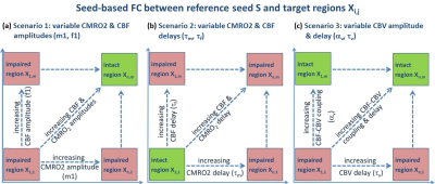
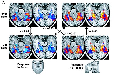
Regions di er for experimental groups (for example { healthy controls versus individuals with mild cognitive impairment)?Similar to conventional taskrelated fMRI, the BOLD fMRI signal is measured throughout the experiment (panel a) Conventional taskdependent fMRI can be used to select a seed region of interest (panel b) To examine the level of functional connectivity between the selected seed voxel i and a second brain region j (for example a region in the


Plos One Probing The Interoceptive Network By Listening To Heartbeats An Fmri Study


Ismrm Digital Posters Page Fmri Connectivity


Concepts And Principles Of Clinical Functional Magnetic Resonance Imaging Chapter 13 The Cambridge Handbook Of Research Methods In Clinical Psychology


Mriquestions Com Uploads 3 4 5 7 Heuvel Reviewbrainnets1 Pdf


Predicting Functional Networks From Region Connectivity Profiles In Task Based Versus Resting State Fmri Data


Resting State Fmri Wikipedia


Fsl Fmri Resting State Seed Based Connectivity Neuroimaging Core 0 1 1 Documentation


Spontaneous Low Frequency Bold Signal Variations From Resting State Fmri Are Decreased In Alzheimer Disease


The Role Of Resting State Functional Mri For Clinical Preoperative Language Mapping Cancer Imaging Full Text


Estimation Of Resting State Functional Connectivity Using Random Subspace Based Partial Correlation A Novel Method For Reducing Global Artifacts Abstract Europe Pmc
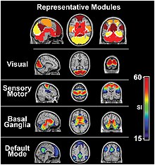


Resting State Fmri Wikipedia


Neuropsychiatric Neurologic Disorder Classification Prediction Brain Signal Processing Lab


Seed Based Connectivity Analysis Of Resting State Fmri Data A Download Scientific Diagram


A Location Of Striatal Seed Regions Green Within Group Functional Download Scientific Diagram


Ieeexplore Ieee Org Iel7 Pdf


Interactively Generated Resting State Networks Z 4 0 From A Single Download Scientific Diagram


Localized Prediction Of Glutamate From Whole Brain Functional Connectivity Of The Pregenual Anterior Cingulate Cortex Biorxiv


Comparison Of Task Based Fmri Result And Resting State Fmri In A Single Download Scientific Diagram


Changes In Thalamus Connectivity In Mild Cognitive Impairment Evidence From Resting State Fmri European Journal Of Radiology
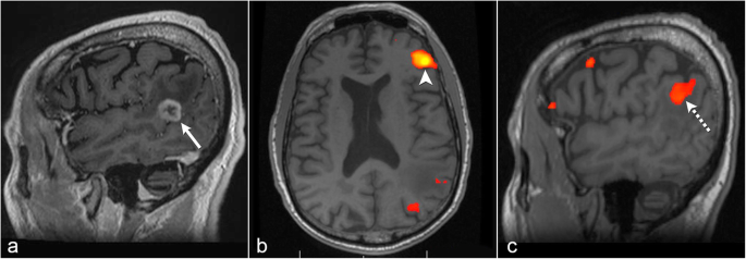


The Role Of Resting State Functional Mri For Clinical Preoperative Language Mapping Cancer Imaging Full Text
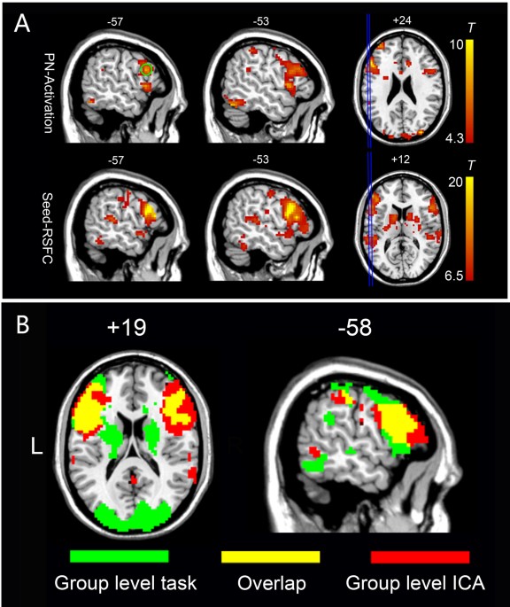


An Automated Method For Identifying An Independent Component Analysis Based Language Related Resting State Network In Brain Tumor Subjects For Surgical Planning Scientific Reports
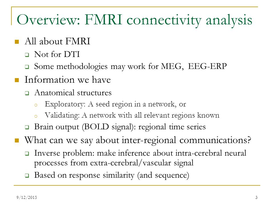


Fmri Connectivity Analysis In Afni Ppt Video Online Download
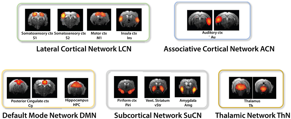


Frontiers Resting State Fmri In Mice Reveals Anesthesia Specific Signatures Of Brain Functional Networks And Their Interactions Frontiers In Neural Circuits


Seed Based Connectivity Analysis Of Resting State Fmri Data A Download Scientific Diagram


Link Springer Com Content Pdf 10 1007 2f978 3 319 567 2 9077 1 Pdf


Resting State Fmri Wikipedia
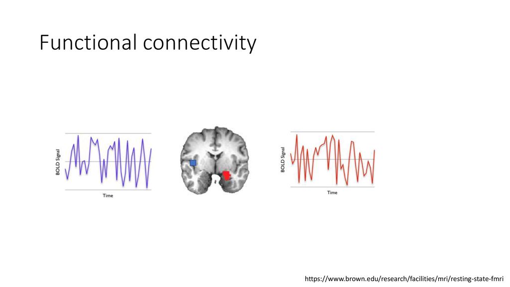


Characterizing The Spectrum Of Task Fmri Connectivity Approaches Ppt Download
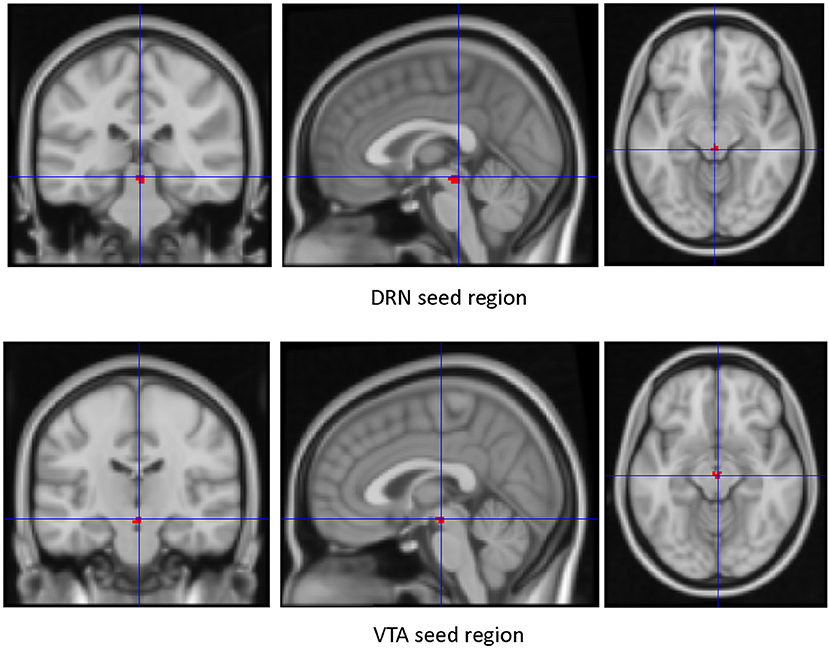


Frontiers Resting State Functional Connectivity Of Dorsal Raphe Nucleus And Ventral Tegmental Area In Medication Free Young Adults With Major Depression Psychiatry


Fmri Peak Activation Group Differences For Each Fc Seed Region Download Table


There Were Significant Fc Fmri Values Relative To The Pcc Seed Region Download Scientific Diagram


High Amplitude Cofluctuations In Cortical Activity Drive Functional Connectivity Pnas


A Window Into The Brain Advances In Psychiatric Fmri


Cancerimagingjournal Biomedcentral Com Track Pdf 10 1186 S 0 W Pdf
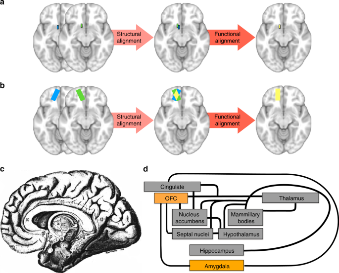


An Improved Neuroanatomical Model Of The Default Mode Network Reconciles Previous Neuroimaging And Neuropathological Findings Communications Biology


Rsfc Correlations Seed Regions Consisted Of The 5 Functional And 2 A Download Scientific Diagram


Defining The Seed Region Of Interest Ppi Analysis Investigates Download Scientific Diagram
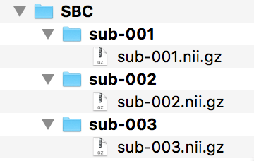


Fsl Fmri Resting State Seed Based Connectivity Neuroimaging Core 0 1 1 Documentation


Figure 3 From A Window Into The Brain Advances In Psychiatric Fmri Semantic Scholar
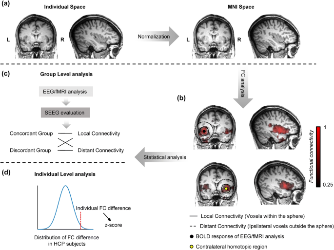


Enhanced Regional Functional Connectivity Indicates Seizure Onset Zone Springerlink
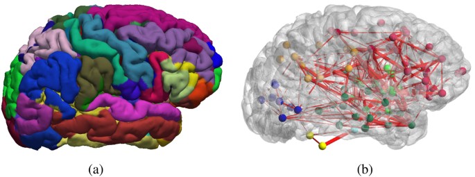


Function Specific And Enhanced Brain Structural Connectivity Mapping Via Joint Modeling Of Diffusion And Functional Mri Scientific Reports


Effects Of Isoflurane Anesthesia On Resting State Fmri Signals And Functional Connectivity Within Primary Somatosensory Cortex Of Monkeys Wu 16 Brain And Behavior Wiley Online Library


Figure 4 From Eeg Alpha Power Modulation Of Fmri Resting State Connectivity Semantic Scholar


Fmri Results A Cortical Areas More Strongly Activated During Mi Download Scientific Diagram


Resting State Functional Mri Everything That Nonexperts Have Always Wanted To Know American Journal Of Neuroradiology


Presurgical Resting State Functional Mri Language Mapping With Seed Selection Guided By Regional Homogeneity Hsu Magnetic Resonance In Medicine Wiley Online Library


Assessing Functional Connectivity In The Human Brain By Fmri Abstract Europe Pmc


Http Www Gestaltrevision Be Pdfs Lss14 Dm Leuven 12 Aug 14 Pdf


Plos One Resting State Fmri Reveals Diminished Functional Connectivity In A Mouse Model Of Amyloidosis


Commonly Used Analysis Methods In Functional Mri Studies For Download Scientific Diagram


Resting State Functional Mri Everything That Nonexperts Have Always Wanted To Know American Journal Of Neuroradiology


Active And Passive Fmri For Presurgical Mapping Of Motor And Language Cortex Intechopen


Plos One Unravelling The Intrinsic Functional Organization Of The Human Striatum A Parcellation And Connectivity Study Based On Resting State Fmri


Seed Regions Of The Seed Based Resting State Analysis Seed Regions And Download Scientific Diagram


Predicting Functional Networks From Region Connectivity Profiles In Task Based Versus Resting State Fmri Data


Multimodal Neuroimaging Of Prosody Applied Social Neuroscience And Perception Laboratory
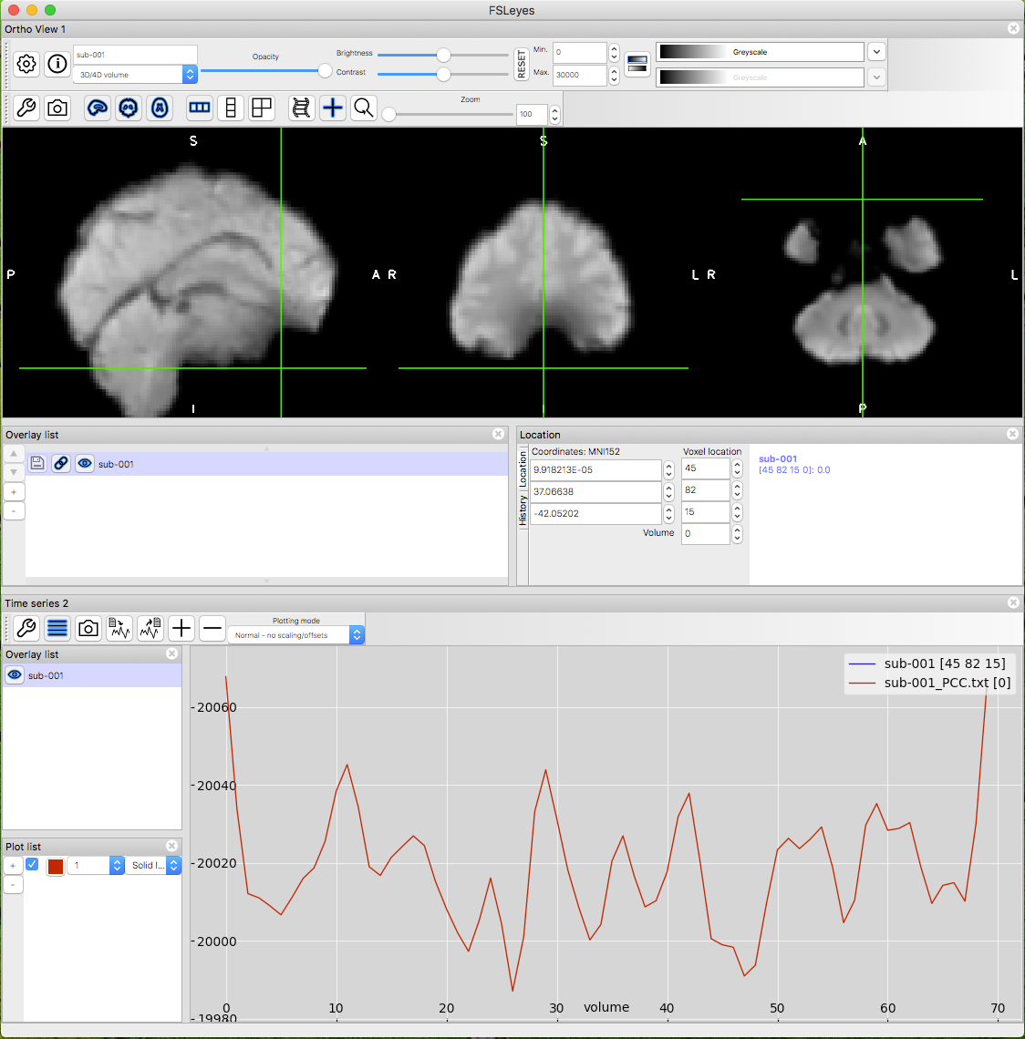


Fsl Fmri Resting State Seed Based Connectivity Neuroimaging Core 0 1 1 Documentation


Plos One Resting State Fmri Reveals Diminished Functional Connectivity In A Mouse Model Of Amyloidosis


Patient Tailored Connectivity Based Forecasts Of Spreading Brain Atrophy Sciencedirect


Seed Based Resting State Fmri Functional Connectivity Matrices


Defining The Seed Region Of Interest Ppi Analysis Investigates Download Scientific Diagram


Fsl Fmri Resting State Seed Based Connectivity Neuroimaging Core 0 1 1 Documentation


1


Changes In Resting State Functional Brain Activity Are Associated With Waning Cognitive Functions In Hiv Infected Children Abstract Europe Pmc


Fsl Fmri Resting State Seed Based Connectivity Neuroimaging Core 0 1 1 Documentation


Http Www Gestaltrevision Be Pdfs Lss14 Dm Leuven 12 Aug 14 Pdf


The Human Brain Is Intrinsically Organized Into Dynamic Anticorrelated Functional Networks Pnas


Figure 3 From Localization Of Broca S And Wernicke S Areas With Resting State Fmri Semantic Scholar


The Role Of Resting State Functional Mri For Clinical Preoperative Language Mapping Cancer Imaging Full Text


Plos One Coupled Intrinsic Connectivity Distribution Analysis A Method For Exploratory Connectivity Analysis Of Paired Fmri Data


1


Identifying Resting State Networks From Fmri Data Using Icas By Gili Karni Towards Data Science


Resting State Functional Mri Everything That Nonexperts Have Always Wanted To Know American Journal Of Neuroradiology


Traditional Seed Based Analysis Approach Download Scientific Diagram
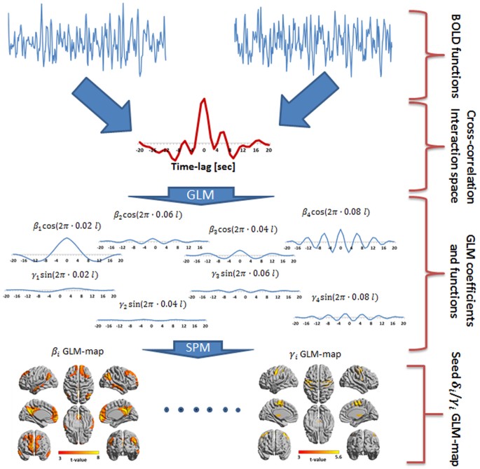


Frequency Phase Analysis Of Resting State Functional Mri Scientific Reports


Resting State Functional Mri Everything That Nonexperts Have Always Wanted To Know American Journal Of Neuroradiology


Does Resting State Functional Connectivity Differ Between Mild Cognitive Impairment And Early Alzheimer S Dementia Journal Of The Neurological Sciences


Resting State Fmri Wikipedia
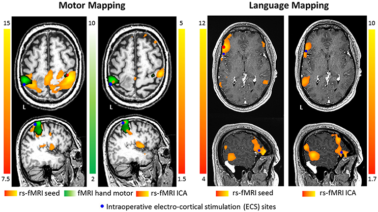


Frontiers Pre Surgical Brain Mapping To Rest Or Not To Rest Neurology


0 件のコメント:
コメントを投稿